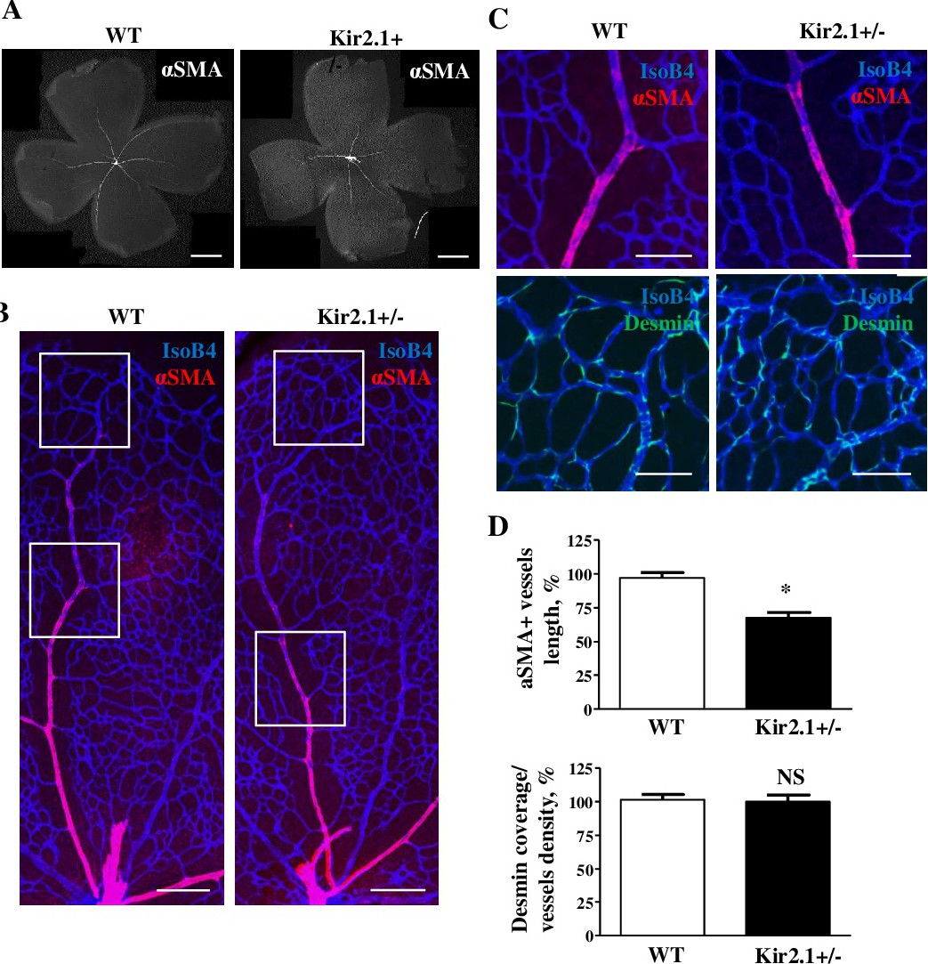
Fig. 3. Deficiency of Kir2.1 decreased the smooth muscle cell coverage but does not change pericytes coverage of the retinal vasculature. (A) Mouse retinas were stained with alfa smooth muscle actin (αSMA) to visualize vSMCs. Low magnification images of retina showed decreased the αSMA-positive vessel length in Kir2.1+/- mice compare to wild type. High magnification images of mouse retina stained with αSMA (red) and Isolectin B4 (IsoB4, blue) showed the decreased length of αSMA-covered artery. Panel 3C shows the magnification of the inserts, indicated as squares in Panel 3B. The two upper squares of Panel B are expanded to show Desmin and the two lower squares in Panel B are expanded to show αSMA. Please note that the αSMA region chosen for WT mice is much closer to the periphery of the retina than the region chosen for Kir2.1+/- mice. This is consistent with a decrease in smooth muscle coverage. (D) Quantification of the αSMA and desmin coverage are shown in lower panel. Data are means ± SD of at least six mice per group. *P˂0.05. Scale bar panel A,B,C: 100 µm.